Mitosis Under Microscope Biology Diagrams Learn the steps of mitosis and what to look for under a microscope in each phase. See examples of prophase, metaphase, anaphase and telophase slides and how to prepare them.
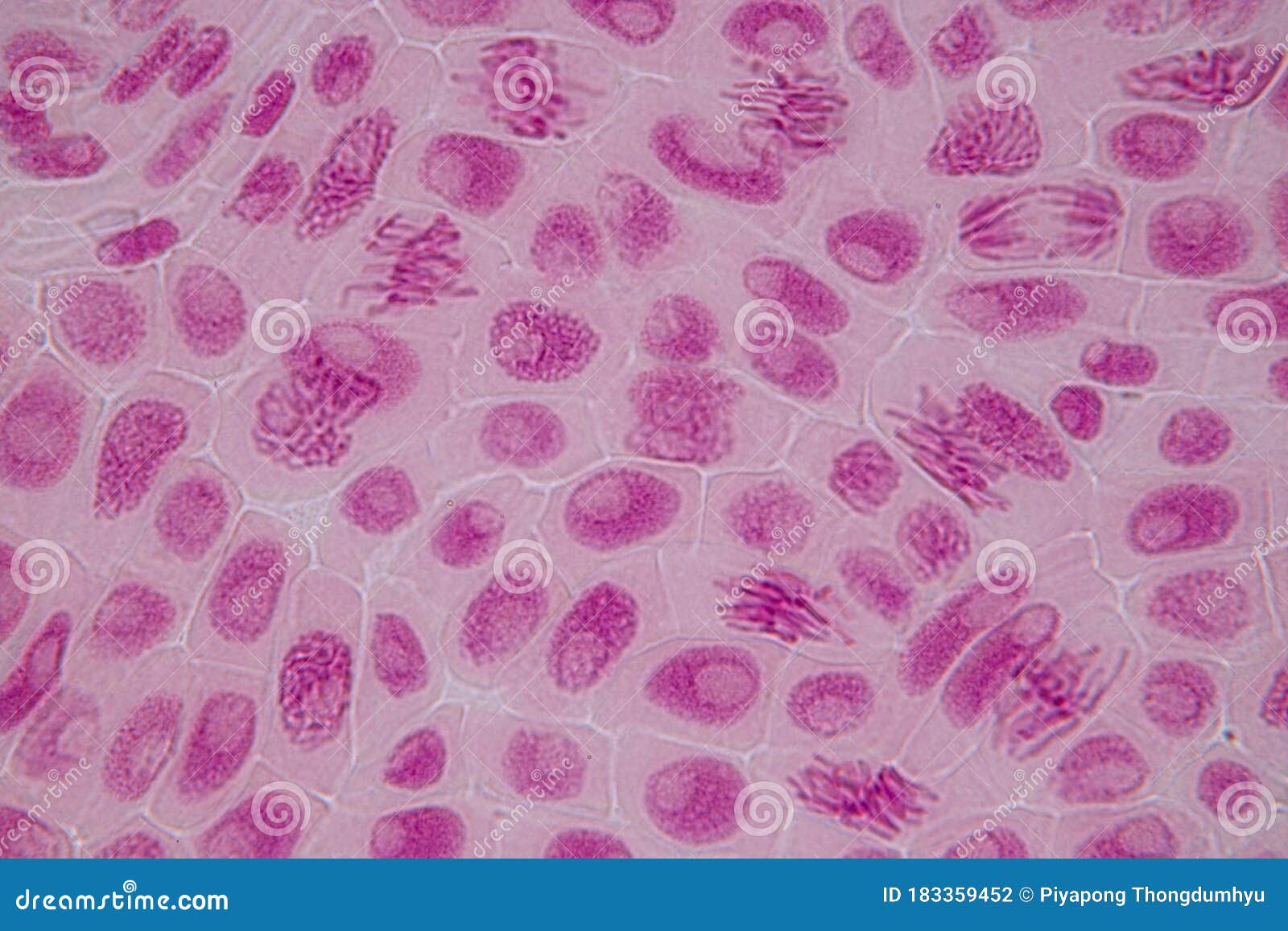
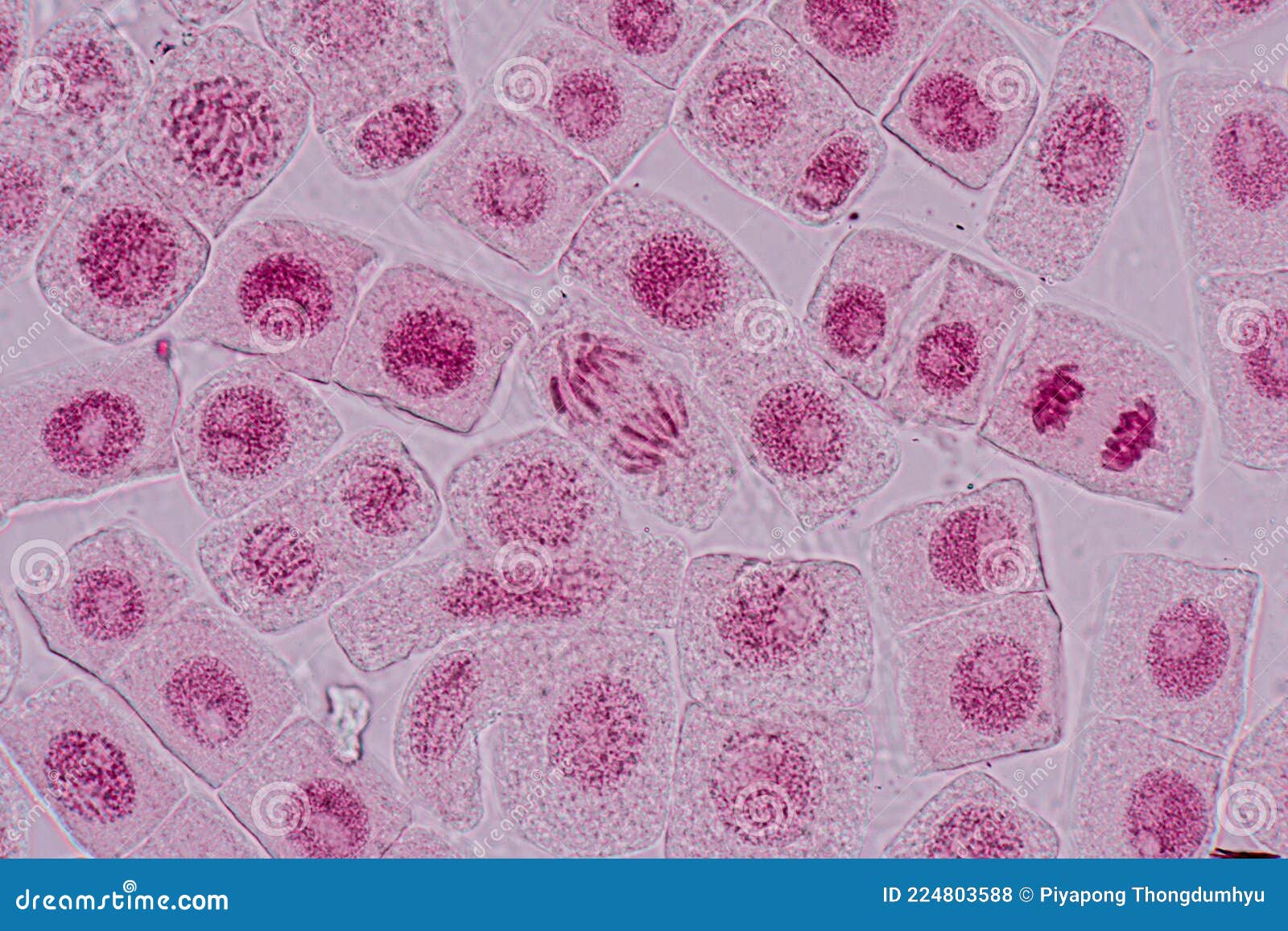
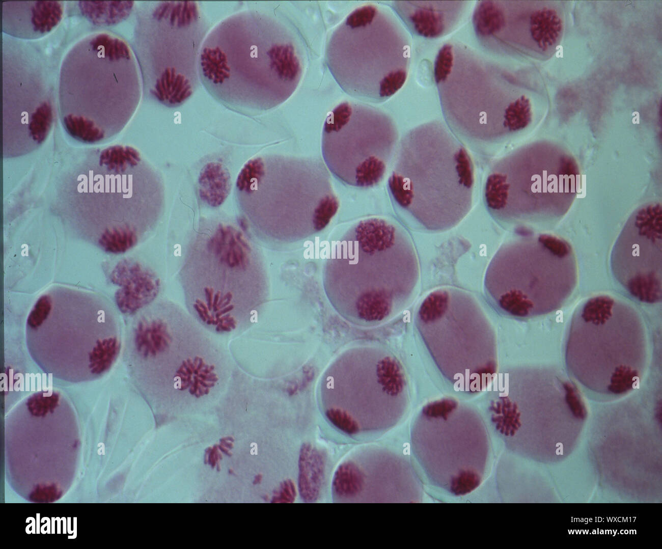
View the cells under a microscope. Analysis. Cells undergoing mitosis (similar to those in the images below) can be seen and drawn. Annotations can then be added to these drawings to show the different stages of mitosis. Limitations. The preparation of tissue for microscope slides can damage cells and alter their appearance

What Do the Stages of Mitosis Look Like Under a Microscope? (Images ... Biology Diagrams
This video takes you through microscope images of cells going through mitosis and identifies the different phases under the microscope and on a micrograph.
Learn the steps of mitosis with photomicrographs and a Java tutorial. See how chromosomes condense, align, separate and divide in onion root tips. Mitosis in an animal cell. Cells from the Chinese Hamster Ovary are shown undergoing mitosis. Beginning with a cell spread on the substrate, follow prophase,
Recognising the Stages of Mitosis Biology Diagrams
Learn about the process of mitosis, or cell division, and see drawings and pictures of the four phases. Use a microscope and a stained onion root tip slide to observe the chromosomes in different cells.